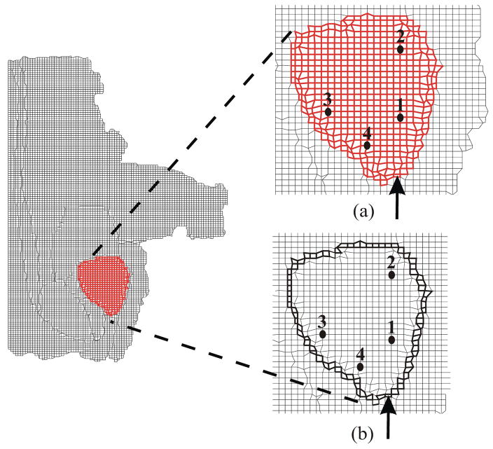
Fig. 6.
FE mesh of the prostate with its surrounding tissue generated from the segmented MR image used for simulation studies. (a) Close-up view of the simulation case where no cohesive elements surround the prostate (NoCoh). (b) Close-up view of the simulation case where prostate gland is surrounded by cohesive elements (in bold black) (Coh). Also shown are the data points at which nodal displacement were measured and the point of applied displacement used to simulate needle insertion.

