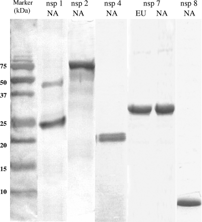
FIG. 1.
Sodium dodecyl sulfate-polyacrylamide gel electrophoresis of recombinant PRRSV nsp preparations, followed by Coomassie blue staining. The left lane shows the protein molecular mass standard; the remaining lanes represent nsp1, nsp2, nsp4, nsp7, and nsp8 preparations, as indicated. NA, North American genotype (type II); EU, European genotype (type I). Note that nsp1 is further cleaved into nsp1α and nsp1β subunits (6, 13). Intact nsp1 and 26-kDa nsp1β eluted from the immobilized metal affinity column are shown in the second lane from the left.

