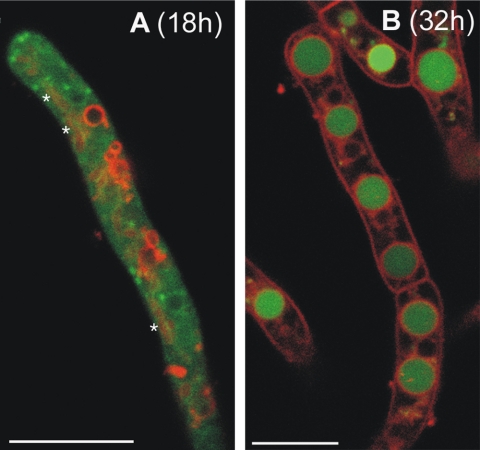
FIG. 2.
Age-dependent distribution of RaVC in living hyphae. The hyphae grown in liquid culture were imaged by confocal microscopy either 18 (A) or 32 h (B) after inoculation. The two figures are of merged images showing pH-dependent RaVC fluorescence (green) and the localization of membrane-selective probe SynaptoRed 2 fluorescence (red). Note that RaVC is localized in the cytoplasm in 18-h-old hyphae (A) and is mostly localized in large putative vacuoles in 32-h-old hyphae (B). Tubular vacuoles in panel A are indicated by asterisks; they are not as clear as the labeled membranes of the spherical vacuoles. Bars = 7.5 (A) and 10 μm (B).

