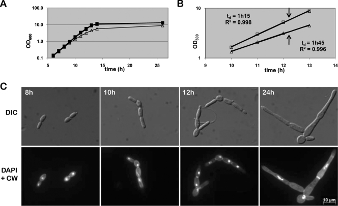
FIG. 3.
Cdc53p functions as a repressor of filament development. (A) Growth curve of strains KTY3 (squares) and KTY31 (triangles) in liquid YPD. Closed shapes indicate growth in the absence of doxycycline, whereas open shapes represent growth in the presence of doxycycline. Due to superimposition of the three growth curves of KTY3 with or without doxycycline and KTY31 without doxycycline, only the closed squares of KTY3 without doxycycline can be seen. (B) Determination of exponential growth phase and growth rate of the control strain KTY3 (squares) and the cdc53 mutant strain KTY31 (triangles), both in the presence of doxycycline. The coefficients of determination (R2) were calculated using the R-square function of Excel. The arrows indicate the time point of cell harvest of both strains for microarray analysis. (C) Time course microscopic analysis of cells of strain KTY31 under Cdc53p-depleting conditions. Calcofluor white and DAPI staining of exponential grown cells display septa at mother bud necks and at further constrictions, indicating pseudohyphal growth mode. Stationary-phase cells (24 h) are more elongated, with location of septa indicating a mix of pseudohyphae and hyphae.

