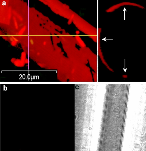
FIG. 5.
(a) Glass wool exposed for 30 min to an exponentially growing B. cereus culture before removal and staining with propidium iodide, viewed by LSCM. The left panel is a composite x-y image, and the right panel is a y-z image of the in silico preparation at the position of the line. (b and c) Glass wool exposed to sterile LB broth for 2 h and then stained with propidium iodide and viewed by LSCM in the red channel (b) and by differential interference contrast microscopy (c).

