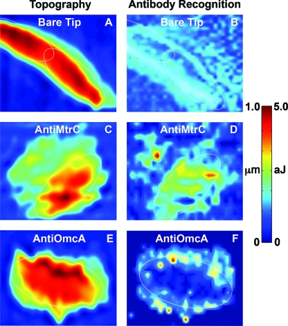
FIG. 3.
Ig-RFM of live S. oneidensis MR-1 cells deposited on a hematite (α-Fe2O3) thin film. Height image (A) and corresponding Ig-RFM image (B) for a bare unfunctionalized Si3N4 tip. Height and corresponding Ig-RFM image for a tip functionalized with anti-MtrC (C and D) or anti-OmcA (E and F). Each panel contains a thin white oval showing the approximate location of the bacterium on the hematite surface. A color-coded scale bar is shown on the right (height in micrometers [μm], and the work required to separate the tip from the surface in attojoules [aJ]).

