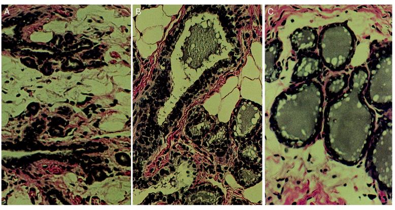
Figure 1.
(A) Histological section of the mammary gland of an untreated 12-week-old virgin Lewis rat. Note the presence of empty ducts with small alveolar buds. (B) Histological section of the differentiated mammary gland of a virgin rat treated for 3 weeks with 30 mg of estradiol and 30 mg of progesterone in silastic capsules. Note the presence of a dilated duct and alveoli filled with secretion and fat droplets. (C) Histological section of the differentiated mammary gland of a virgin rat treated for 3 weeks with 5 mg/kg daily perphenazine. Note the secretion filled mammary ducts and alveoli. (×200.)

