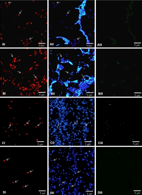
FIG. 2.
Detection of C. sakazakii ATCC 29544 in suspension with a mixed population using SakPNA971. (A) Reconstituted PIF with C. sakazakii and B. cereus. (B) Reconstituted PIF with C. sakazakii, B. cereus, P. aeruginosa ATCC 10145, and S. enterica serotype Enteritidis ATCC 13076. (C) Reconstituted PIF with C. sakazakii, S. enterica serotype Enteritidis ATCC 13076 (100-fold concentrated), and P. aeruginosa ATCC 10145 (100-fold concentrated). (D) Reconstituted PIF with C. sakazakii and S. enterica serotype Enteritidis ATCC 13076 (100-fold concentrated). (Panels I) Detection of C. sakazakii using the red fluorescent SakPNA971 probe. (Panels II) Counterstaining with DAPI (total population). (Panels III) Visualization of the same microscopic field with the green channel (negative control). The arrows indicate C. sakazakii cells that could easily be visualized in the DAPI channel with the red-labeled SakPNA971 probe. All images were obtained using the same exposure time.

