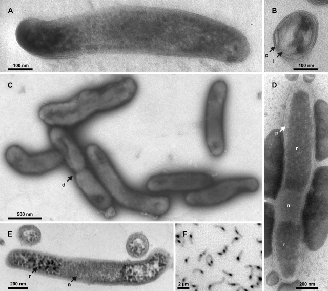
FIG. 1.
Images of strain Pei191T. (A) TEM image of a longitudinal section of a smaller cell, showing both cell poles delimited by the outer membrane. (B) TEM image of a radial section resolving both inner and outer membranes and several ribosomes. (C) Negative-contrast TEM image of cells in the mid-exponential growth phase prepared on a hydrophilized grid. (D) Negative-contrast TEM image of a cell prepared on a nonhydrophilized grid. (E) TEM image of longitudinal and radial sections of larger cells. (F) Fluorescence photomicrograph (negative image) of DAPI-stained cells in the mid-exponential growth phase. Arrows denote cell division (d), inner membrane (i), nucleoid (n), outer membrane (o), periplasmic space (p), and riboplasm (r).

