FIG. 2.
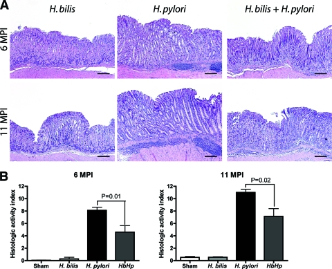
Gastric histology. (A) Representative histopathology of gastric tissues from mice infected with H. bilis or H. pylori or coinfected with H. bilis and H. pylori at 6 to 11 mpi. Lesions were characterized by lymphocyte-predominant mucosal and submucosal infiltrates, multifocal surface erosions and glandular ectasia, oxyntic atrophy, hyperplasia, pseudopyloric metaplasia, and dysplasia. (B) Gastric HAI. Tissues from mice infected with H. pylori, H. bilis, or both H. bilis and H. pylori (n = 15 for all groups) were graded for inflammation, epithelial defects, atrophy, hyperplasia, pseudopyloric metaplasia, dysplasia, hyalinosis, and mucous metaplasia. A gastric HAI was generated by combining scores for all criteria except hyalinosis and mucous metaplasia, which may develop irrespective of helicobacter infection. The HAI was higher in H. pylori-infected mice compared to H. bilis/H. pylori-infected mice (6 mpi, P < 0.01; 11 mpi, P < 0.02).

