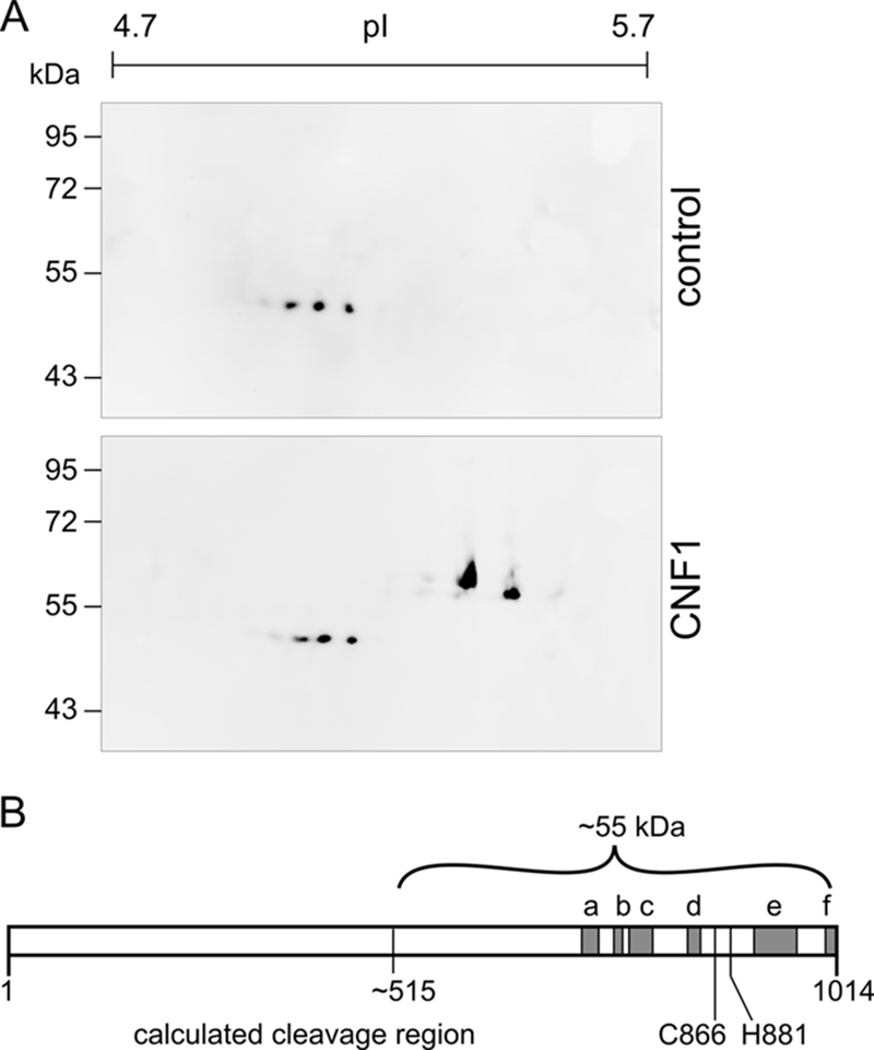
FIG. 3.

Immunoprecipitation of the CNF1 fragment. HeLa cells (109 cells) were incubated with CNF1 (1 μg/ml) or without the toxin overnight and lysed. Following fractionation of the lysates, cytosols were prepared, and immunoprecipitation with a monoclonal antibody against the C terminus of CNF1 was performed. Part of the precipitate (75%) was separated by 2D gel electrophoresis and stained with Coomassie blue, and the corresponding spot (not present in the control) was prepared for MALDI-TOF analysis. Only peptides with an error between −20 and 20 were considered. The positions of identified peptides which include amino acids 693 to 710 (a), 732 to 741 (b), 753 to 775 (c), 821 to 832 (d), 900 to 944 (e), and 990 to 1012 (f) are indicated by gray boxes in panel B. The estimated cleavage region is located around amino acid 515 (55 kDa, ∼500 amino acids). Part of each precipitate (25%) was separated by 2D gel electrophoresis, and the CNF1 fragment was detected by Western blotting (A).
