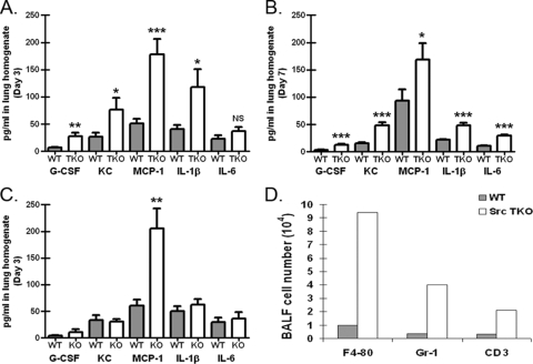
FIG. 3.
Increased lung proinflammatory response in P. murina-exposed Hck−/− Fgr−/− Lyn−/− mice. C57BL/6 (WT) and Hck−/− Fgr−/− Lyn−/− (TKO) mice were inoculated with 2 × 10⁵ P. murina cysts via the intratracheal route. (A and B) At 3 days (A) and 7 days (B) postinoculation, lungs were collected, and clarified supernatants from lung homogenates were analyzed for G-CSF, CXCL1/KC, CCL2/MCP-1, IL-1β, and IL-6 levels by Bio-Plex. The data are cumulative data from three independent studies with five mice per group. The bars and error bars indicate the means and standard errors of the means. * and ***, P < 0.05 and P <0.001, respectively (unpaired two-tailed Student's t test). (C) Three days postinoculation, lungs were collected from Lyn−/− mice, and clarified supernatants from lung homogenates were analyzed for G-CSF, CXCL1/KC, CCL2/MCP-1, IL-1β, and IL-6 levels by Bio-Plex. The data are cumulative data from three independent studies with five mice per group. The bars and error bars indicate the means and standard errors of the means. **, P < 0.01 (unpaired two-tailed Student's t test). (D) C57BL/6 and Hck−/− Fgr−/− Lyn−/− (Src TKO) mice were inoculated with 2 × 10⁵ P. murina cysts via the intratracheal route. Three days postinoculation, bronchoalveolar lavage was performed, and cells were stained for F4/80 (macrophages), Gr-1 (neutrophils), and CD3 (T cells) and assessed by flow cytometry. The data are representative data from one of two independent studies. BALF, bronchoalveolar lavage fluid.

