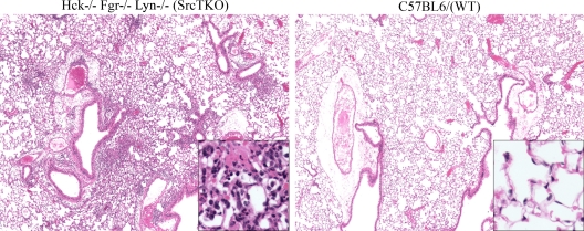
FIG. 4.
Histological evidence for inflammatory differences in the lungs of P. murina-exposed Hck−/− Fgr−/− Lyn−/− mice: representative hematoxylin- and eosin-stained lung sections from (A) Hck−/− Fgr−/− Lyn−/− (Src TKO) mice and (B) wild-type (WT) mice challenged intratracheally with 2 × 10⁵ P. murina cysts for 3 days. Original magnification, ×40. (Insets) Magnification, ×200.

