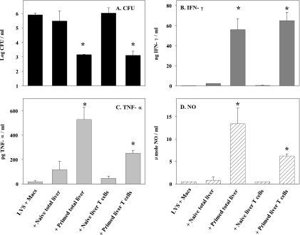
FIG. 5.
LVS-immune liver T lymphocytes control intramacrophage Francisella growth. Confluent macrophages derived from bone marrow of WT C57BL/6J were infected at an MOI of 1:40 with LVS and cocultured with the indicated naïve or LVS-immune total or T-cell-enriched populations. Naïve splenic or liver lymphocytes were obtained from WT C57BL/6 mice, and primed lymphocytes (both liver and spleen) were obtained from C57BL/6J mice given 105 LVS i.d. 1.5 months earlier, using 1 × 10⁶ added lymphocytes for all 48-well cocultures. In this experiment, enriched T-cell populations were 92% CD3+ TCRβ+, 4% CD11b+ CD11c−, 3.5% CD11b+ CD11c+, 0.7% NK1.1+ DX5+, and <0.5% CD19+. Numbers of CFU ± SEM were determined on day 3 from cultures containing only LVS-infected macrophages (LVS + Macs) or containing LVS-infected macrophages cocultured with the indicated cell populations and antibodies. Supernatants from the corresponding cultures were obtained prior to macrophage lysis and assessed for quantities of IFN-γ (B), TNF-α (C), or nitric oxide (D). *, P < 0.05 compared to the corresponding cultures of LVS-infected macrophages without added lymphocytes. The figure depicts one representative experiment of four total experiments of similar design and outcome.

