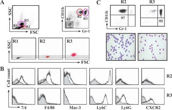
FIG. 3.
In the livers of L. monocytogenes-infected mice, the CD11b+ Gr-1int cells are monocytes and CD11b+ Gr-1hi cells are neutrophils. Mice were infected with 2 × 10⁴ L. monocytogenes bacteria and injected with 100 μg of 3E1. At 16 h p.i., HILs were isolated, stained with antibodies, and analyzed by FACS. (A) Cells were double stained with PE-conjugated anti-CD11b and FITC-conjugated anti-Gr-1 antibodies and gated on CD11b+ Gr-1int (R2) and CD11b+ Gr-1hi (R3). R2 and R3 cells were further analyzed by SSC and forward scatter (FSC) parameters. (B) Expression of surface markers on CD11b+ Gr-1int cells and CD11b+ Gr-1hi cells. HILs were triple stained with antibodies for CD11b, Gr-1, and each surface marker. After being gated on CD11b+ Gr-1int (R2) and CD11b+ Gr-1hi (R3) as in panel A, R2 and R3 cells were further analyzed for the expression of 7/4, F4/80, Mac-3, Ly6C, Ly6G, or CXCR2. (C) Nuclear morphology reveals that CD11b+ Gr-1int cells are monocytes and CD11b+ Gr-1hi cells are neutrophils. The R2 and R3 cells in panel A were sorted by FACS, cytospun, and stained with Diff-Quik. The top row shows the purity of the sorted cells, and bottom row shows photomicrographs (magnification, ×1,000). The data are representative of experiments repeated three times with four mice per group.

