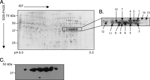
FIG. 4.
Two-dimensional SDS-PAGE and Western blot analyses of A. phagocytophilum NCH-1A Msp2(P44) proteins. Hydrophobic pellet fractions enriched for A. phagocytophilum NCH-1A outer membrane proteins were isoelectric focused (IEF) in IPG strips (pH 5.0 to 8.0), which was followed by resolution in the second dimension in SDS-polyacrylamide (4 to 20%) gels. (A) Silver-stained gel. Msp2(P44) proteins that were excised for LC-MS/MS identification and detected by MAb 20B4 are indicated by a box. (B) Enlarged view of the region of the silver-stained gel in panel A indicated by the box. Numbered spots were excised and identified by LC-MS/MS. (C) MAb 20B4 recognized spots in the predicted size range for full-length Msp2(P44) proteins.

