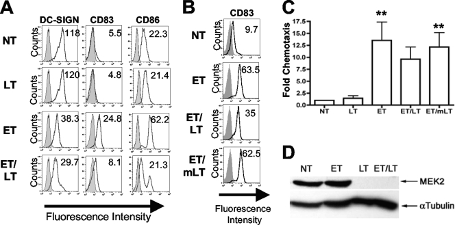
FIG. 3.
Effects of LT on ET-mediated DC maturation and chemotaxis. (A and B) MDDCs were pretreated with 10 μg/ml polymyxin B for 45 min and were then treated either with 100 ng/ml PA plus 100 ng/ml EF (ET), 100 ng/ml PA plus 100 ng/ml LF (LT), 100 ng/ml PA plus 100 ng/ml EF plus 100 ng/ml LF (ET/LT), or no treatment (NT) (A) or with 100 ng/ml PA plus 100 ng/ml EF (ET), 100 ng/ml PA plus 100 ng/ml EF plus 100 ng/ml LF (ET/LT), 100 ng/ml PA plus 100 ng/ml EF plus 100 ng/ml LF(H719C) (ET/mLT), or no treatment (NT) (B). Cells were harvested 48 h after treatment and incubated with anti-DC-SIGN-FITC, anti-CD83-APC, and anti-CD86-FITC and then subjected to flow cytometric analysis. The data depicted represent the results for one of five independent experiments conducted on MDDCs generated from five different donors. The histograms depict data for the unstained sample (shaded histogram), data for the stained sample (solid black line), and the geometric mean fluorescence under each treatment condition. (C) MDDCs were pretreated with 10 μg/ml polymyxin B for 45 min and then treated with either 100 ng/ml PA plus 100 ng/ml EF (ET), 100 ng/ml PA plus 100 ng/ml LF (LT), 100 ng/ml PA plus 100 ng/ml EF plus 100 ng/ml LF (ET/LT), or 100 ng/ml PA plus 100 ng/ml EF plus 100 ng/ml LF(H719C) (ET/mLT). Following 48 h of treatment, the migration of MDDCs toward MIP-3β was measured using a transwell assay employing uncoated inserts (5-μm pore). Data are shown as the increase in migration compared with that of immature (NT) MDDCs for each donor and are the average values for five donors ± SD. Statistical significance was calculated using one-way ANOVA with Bonferroni's multiple comparison test. **, P < 0.05, compared to NT. (D) Cells were pretreated with 10 μg/ml polymyxin B for 45 min and then treated for 2 h with 500 ng/ml PA plus 500 ng/ml EF (ET), 500 ng/ml PA plus 500 ng/ml LF (LT), or 500 ng/ml PA plus 500 ng/ml EF plus 500 ng/ml LF (ET/LT) or were left untreated (NT). WCEs (20 μg/sample) were analyzed by Western blotting using antibodies specific for MEK2 (N terminus) or α-tubulin.

