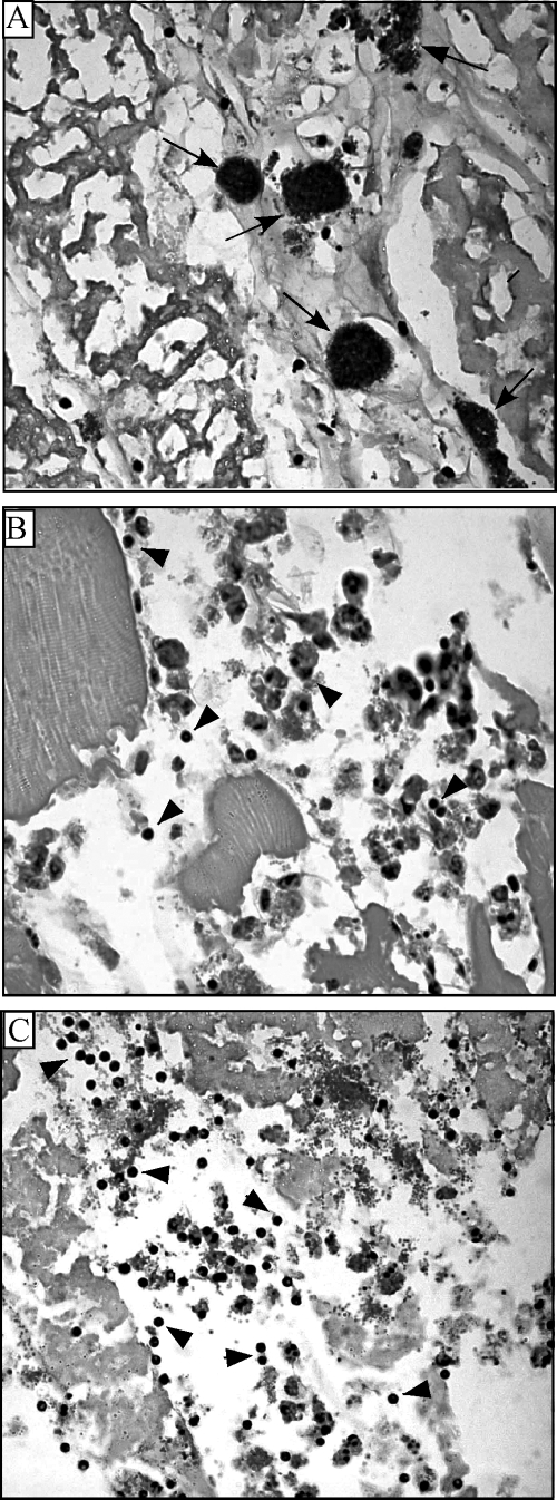
FIG. 2.
Zebrafish histology after S. pyogenes infection. Tissue sections were prepared from the dorsal muscle 24 h postinfection with 105 CFU wild-type bacteria (A), a silB mutant (B), and a silC mutant (C) and stained with hematoxylin and eosin. The arrows in panel A point to aggregates of bacteria, and the arrowheads in panels B and C point to inflammatory cells at the site of infection. Magnification, ×1,000.

