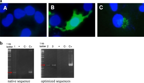
FIG. 1.
(a) Immunofluorescence analysis of B16 melanoma cells transfected with either pVax-FcBoNT/A (A), pVax-Fc*BoNT/A (B), or pVax-SecEpo-Fc*BoNT/A (C), stained as described in Materials and Methods, and visualized by fluorescence microscopy. (b) RT-PCR of FcBoNT/A, Fc*BoNT/A, and SecEpo-Fc*BoNT/A mRNAs 48 h after transfection of B16 cells. Lane 1, FcBoNT/A; lane 2, Fc*BoNT/A; lane 3, SecEpo-Fc*BoNT/A; lanes −, RNA alone; lanes C−, control PCR on water; lanes C+, control PCR on plasmid. Specific primers used were as follows: for FcBoNT/A (259-bp product), 5′-TGCATCACAGGCAGGCGTAG-3′ (forward) and 5′-CCCATGAGCAACCCAAAGTCC-3′; and for Fc*BoNT/A and SecEpo-Fc*BoNT/A (703-bp product), 5′-GCCTGAACTACGGCGAGATCATCTGG-3′ (forward) and 5′-GATCTCCAGGGCGCTCAGGATCTT-3′ (reverse). The red arrows indicate the expected band size (259 bp or 703 bp).

