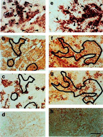
Figure 3.
(a) In situ hybridization of antisense probe GfA 1a (dark purple staining) to the proximal pars distalis of the goldfish pituitary. To demonstrate the overlap of areas of hybridization with gonadotrophs and somatotrophs, selected areas (a) of hybridization (dark purple stain) indicated by arrowheads are projected onto adjacent sections immunocytochemically stained for GtH-II (brown stain; Fig. b) and growth hormone (brown stain; c). (d) Negative in situ hybridization reaction of sense probe to GfA to the goldfish pituitary. (e) In situ hybridization of antisense probe GfB1a (dark purple staining) to the goldfish pituitary. To demonstrate the overlap of areas of hybridization with gonadotrophs and somatotrophs, a selected area (e) of hybridization (dark purple stain) indicated by arrowheads is projected onto adjacent sections immunocytochemically stained for GtH-II (brown stain; f) and growth hormone (brown stain; g). (h) Negative in situ hybridization reaction of sense probe to GfB to the goldfish pituitary. (a–d) Female goldfish undergoing ovarian recrudescence, gonadosomatic index = 4.1%. (e–h) Mature male goldfish, gonadosomatic index = 2.3%. (Bar in h = 100 μm; all panels are the same magnification.)

