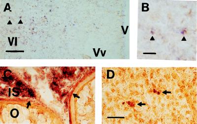
Figure 4.
(a) In situ hybridization of antisense probe GfA 2a (dark purple staining) to perikarya in the area ventralis telencephali of mature male goldfish, gonadosomatic index = 2.2%. Arrowheads indicate two perikarya shown at higher magnification in b. (Bar in a = 100 μm; bar in b = 20 μm.) (c) In situ hybridization of antisense probe GfA 1a (dark purple staining) to interstitial tissue (IS) of the goldfish ovary and the theca cell layer surrounding oocytes (arrowheads). (d) In situ hybridization of antisense probe GfA 1a (dark purple staining) to scattered hepatocytes (arrowheads) in the goldfish liver. (Bar in d = 20 μm; panels c–d are all the same magnification.) (c) Female goldfish, gonadosomatic index = 4.1%. (d) Male goldfish, gonadosomatic index = 2.3%. V, brain ventricle; Vl, area ventralis telencephali pars lateralis; Vv, area ventralis telencephali pars ventralis; IS, interstitial tissue; O, ovary.

