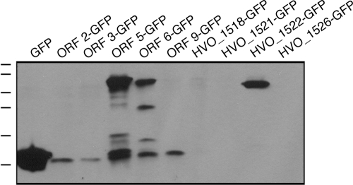
FIG. 3.
The appearance of GFP-containing fusion proteins reveals genes that are expressed. As described in Materials and Methods, H. volcanii cells were transformed with plasmids carrying a sequence of interest fused at its 3′ end to DNA encoding GFP. This construct was preceded by the 200-bp region immediately preceding the sequence of interest. In a control experiment, plasmid pJAM1020 (19), directing the expression of GFP alone, was employed. Expression of the GFP-containing chimeras was then assessed by immunoblot analysis using antibodies against GFP. The positions of molecular mass markers (100, 70, 55, 40, 35, and 25 kDa) are shown on the left.

