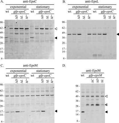
FIG. 2.
Western blot analyses of gfp-epsC and gfp-epsM deletion strains. Cell extracts of log-phase and stationary-phase cultures from the wild-type (wt), gfp-epsC, and ΔepsD (ΔD), ΔepsL (ΔL), and epsM::Tn5 (M−) strains were separated by SDS-PAGE and analyzed by Western blotting with detection by anti-EpsC (A), anti-EpsL (B), and anti-EpsM (C) antisera. (D) Log-phase culture samples of the wt, gfp-epsM, and ΔepsC (ΔC), ΔepsD (ΔD), and ΔepsL (ΔL) strains were immunoblotted with anti-EpsM antisera. Molecular weight markers (in thousands) for all blots are indicated to the left, and the positions of the native proteins and GFP fusions are indicated with black and white triangles, respectively. Full-length GFP-EpsM (white triangle) (D) is partially obscured by a cross-reactive band also present in the wild-type strain. A degradation product of the GFP-EpsM fusion is indicated with a gray triangle (D).

