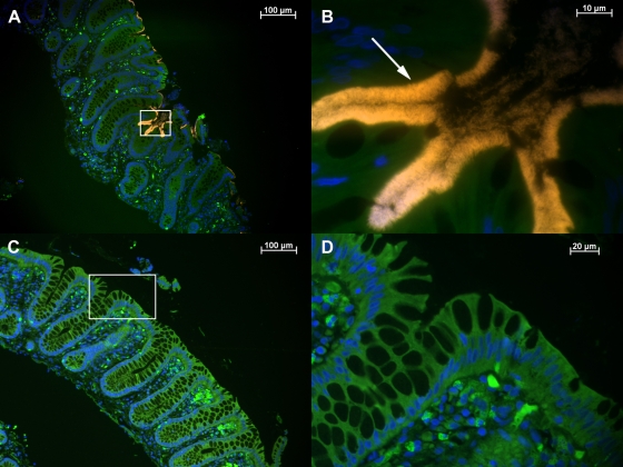
FIG. 3.
Visualization and identification of Brachyspira spp. in a colon biopsy specimen by FISH. Sections of colon biopsy specimens from patient HIS4 (A and B) and control patient 1 (C and D) were hybridized with BRACHYCy3 (orange) and stained with DAPI (blue). (A) Overview. An overlay of the Cy3, fluorescein isothiocyanate, and DAPI filter set results in a bright orange fringe covering the surface epithelium of the crypts and contrasting with the green background fluorescence of the tissue. (B) A higher magnification of the microscopic field boxed in panel A reveals the characteristic end-on attachment (arrow) as well as single Brachyspira sp. organisms in the lumen. (C and D) Absence of spirochetes in normal tissue from a control patient, shown at low and high magnification, respectively.

