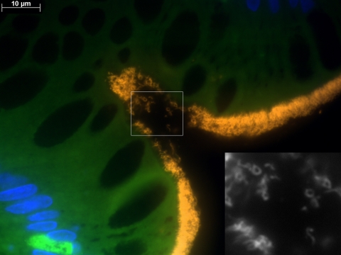
FIG. 4.
Identification of different Brachyspira morphologies by FISH with BRACHY. A section of a colon biopsy specimen from patient HIS4 was hybridized with BRACHYCy3 (orange) and stained with DAPI (blue). In addition to the typical presentation of spirochetes in a helical shape, attached by one end to the surface epithelium, microorganisms with different morphologies can be visualized. Yielding a positive signal with BRACHYCy3, these microorganisms are identified as Brachyspira spp., as shown in the black-and-white image (inset) that was taken with the Cy3 filter set only.

