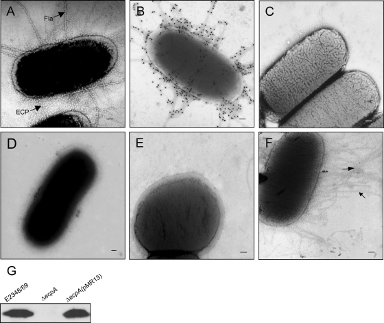
FIG. 1.
ECP on EPEC as shown by electron microscopy and immunogold labeling. (A) Electron micrograph of E2348/69 grown in DMEM displaying abundant peritrichous fine pili (ECP) and flagella (Fla). (B and C) Immunogold labeling of ECP on E2348/69 with anti-ECP antibodies and preimmune serum, respectively. (D) E2348/69 grown in LB medium showing no pili. (E) E2348/69ΔecpA mutant showing no ECP. (F) E2348/69ΔecpA(pMR13) with restored ECP production. Scale bars, 100 nm. (G) Detection of EcpA in normalized HCl-treated whole-cell extracts.

