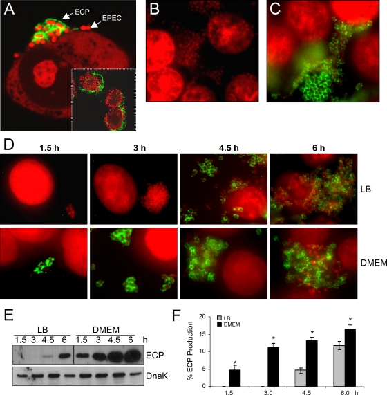
FIG. 2.
ECP on EPEC strains adhering to cultured epithelial cells and kinetics of ECP production. (A) E2348/69 adhering to cultured epithelial cells after 6 h of incubation and reaction with anti-ECP antibodies and Alexa Fluor 488-conjugated secondary antibody (green). Cellular and bacterial DNA was stained with propidium iodide (red). The inset shows a lower magnification of an EPEC microcolony (red) with bacteria producing ECP (green). (B) E2348/69ΔecpA mutant. (C) E2348/69ΔecpA(pMR13). (D) Kinetics of ECP production by bacteria pregrown overnight in LB medium or DMEM. (E) Detection of EcpA in normalized HCl-treated whole-cell extracts. Detection of DnaK with anti-DnaK antibody was used as a loading control. (F) Level of ECP as determined by flow cytometry in bacteria adhering to epithelial cells. The data are representative data from two experiments performed in triplicate. The asterisk indicates that there was a statistically significant difference compared with LB medium-grown bacteria.

