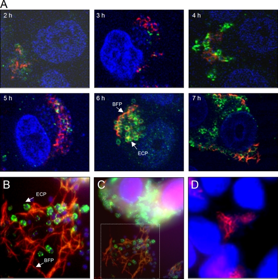
FIG. 4.
Simultaneous production of ECP and BFP during microcolony formation by E2348/69. (A) Kinetics (2 to 7 h) of ECP (green) and BFP (red) expression in the presence of HT-29 cells using confocal microscopy. Cellular and bacterial DNA was stained blue with DAPI. ECP are tightly associated with the bacterial surface, while the BFP extend out from the bacteria throughout the cluster. The confocal microscopy micrographs were taken at a magnification of ×60. (C) IFM detection of ECP and BFP after 6 h of infection. (B) Magnification of the framed area in panel C. Long and thick BFP structures are evident at this time point. (D) E2348/69ΔecpA mutant.

