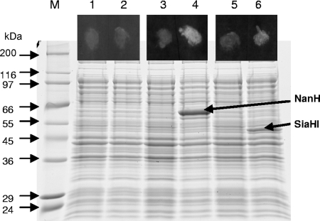
FIG. 2.
Sialidase activity of T. forsythia NanH and SiaHI expressed in E. coli. Strains KCL116 (E. coli pET30; lanes 1 and 2), KCL117 [E. coli BL21(DE3)/pET30::nanH; lanes 3 and 4], and KCL120 [E. coli BL21(DE3)/pET30::siaHI; lanes 5 and 6] were grown in LB until mid-exponential phase and for a further 2 h with (lanes 2, 4, and 6) or without (lanes 1, 3, and 5) induction with 1 mM IPTG. Cells were assayed for sialidase activity in a filter paper spot assay using the fluorogenic substrate 4-MU-NeuNAc (spots in the black boxes above the corresponding lanes). Cell lysates were separated by sodium dodecyl sulfate-10% polyacrylamide gel electrophoresis, and proteins were visualized by Coomassie blue staining.

