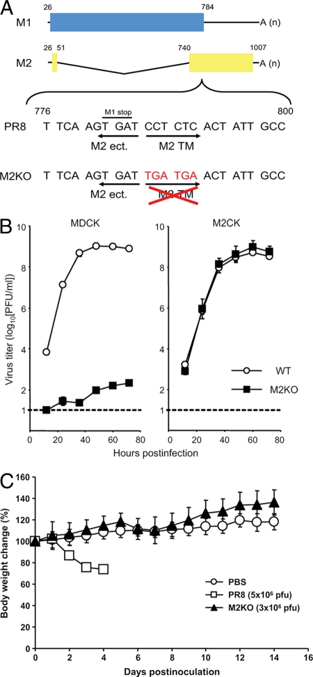
FIG. 1.
Construction of the mutant M segment, growth kinetics of M2KO virus, and change in body weight of mice infected with M2KO virus. (A) Schematic diagram of the mutated M segment of the M2KO virus. Blue and yellow columns represent the open reading frame of the M1 and M2 proteins, respectively. Two stop codons (TGA TGA) were introduced downstream of the open reading frame of the M1 protein in the M segment to eliminate the transmembrane and cytoplasmic tail domains of the M2 protein. M2 ect., M2 TM, and A (n) denote the ectodomain domain of M2, the transmembrane domain of M2, and the poly(A) tail, respectively. Numbers refer to the nucleotide numbers from the 5′ end of the cRNA. (B) Growth properties of PR8 and M2KO viruses in MDCK and M2-expressing MDCK (M2CK) cells. MDCK and M2CK cells were infected with PR8 or M2KO virus at a multiplicity of infection of 0.001. Virus titers in the supernatant of MDCK (left) and M2CK (right) cells at various time points postinfection were determined by using M2CK cells. The dotted line indicates the detection limit of virus titer (10 PFU/ml). (C) Body weight changes in mice inoculated with PBS, PR8, or M2KO virus. The body weights of the control (PBS) or infected (PR8 or M2KO virus) mice were measured daily postinfection. Values are expressed as the mean change in body weight ± standard deviations (n = 8 for PBS and M2KO virus; n = 3 for PR8).

