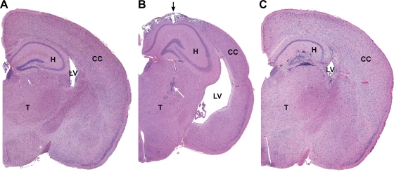
FIG. 4.
Brain from sham-inoculated TG (A) or Ed MeV-infected TG (B) or NT (C) mice, harvested at 28 days p.i. Hydrocephalus in Ed MeV-infected TG mice was associated with a reduction in cerebral cortical (CC) thickness by more than 25%, resulting in the enlargement of the lateral ventricles (LV), whereas hydrocephalic changes were not observed in NT Ed MeV-infected mice surviving to 28 days p.i. These subgross images illustrate the paucity of changes in the hippocampus (H) of TG mice even when there is extensive pannecrosis with mineralization in the overlying cerebral cortex (black arrow) and inflammatory infiltrates (white arrow) of the thalamus (T). Findings for TG mice are contrasted to the more pronounced inflammatory involvement of the hippocampus in an NT mouse, where the degree of involvement of the cerebrum and thalamus is not appreciated at this magnification. The latter brain is from the same animal illustrated in Fig. 5C and D. Sham-inoculated control mice lack microscopic lesions.

