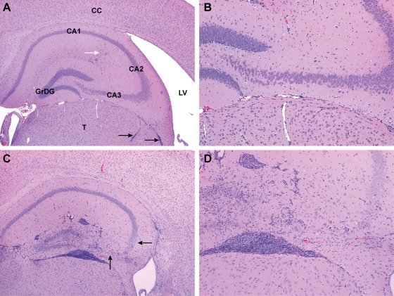
FIG. 5.
Representative pathological anatomical changes in brains of TG (A and B) or NT (C and D) mice at 28 days following intracranial inoculation with Ed MeV. Sections illustrate the hippocampal and dentate gyrus and the adjacent thalamus (T), lateral ventricle (LV), and cerebral cortex (CC), shown here in H&E-stained sections of formalin-fixed tissues at magnification ×4 (A and C) or ×10 (B and D). Granular cell neurons of the dentate gyrus (GrDG) and pyramidal neurons of the CA1, CA2, and CA3 regions of the hippocampus were unaffected in 78% of the TG mice, as shown in panels A and B, where minimal leptomeningeal inflammatory infiltrates (black arrows) and a region of minimal hippocampal gliosis (white arrow) were the only significant findings within these structures. The field illustrated in panel B lacks significant lesions and is indistinguishable from tissue from sham-inoculated controls. The enlarged lateral ventricle is evidence of hydrocephalus, a change observed in 35% of the Ed MeV-infected TG mice. In contrast, pronounced inflammatory and glial responses involving the hippocampal and dentate gyrus were observed in 80% of the NT mice, and there was evidence of pyramidal neuronal necrosis in 56%, illustrated here as a marked reduction in neuronal density in the CA3 region and a moderate reduction in the CA2 region (arrows) (C and D).

