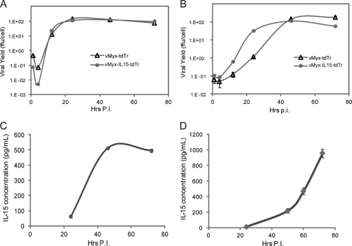
FIG. 2.
Growth kinetics and IL-15 expression from vMyx-IL-15-tdTr. (A) One-step growth of vMyx-IL-15-tdTr in RK-13 cells. RK-13 cells were infected with either vMyx-tdTr or vMyx-IL-15-tdTr at a multiplicity of infection (MOI) of 3. Viral yields from cell lysates were determined at different hours postinfection (Hrs p.i.) in BSC40 cells. (B) One-step growth of vMyx-IL-15-tdTr in murine melanoma B16F10 cells. B16F10 cells were infected with either vMyx-tdTr or vMyx-IL-15-tdTr at an MOI of 3. Viral yields from cell lysates were determined as described in panel A. (C) Concentration of IL-15 in the supernatant of RK-13 cells infected with vMyx-IL-15-tdTr. RK-13 cells were infected at an MOI of 3, and supernatants were collected at 24, 46, and 72 h p.i. for the detection of IL-15 by ELISA (eBioscience) according to the manufacturer's instructions. (D) Concentrations of IL-15 in the supernatant of BGMK cells infected with vMyx-IL-15-tdTr. BGMK cells were infected at an MOI of 10 and supernatants were collected at 24, 50, 60, and 72 h p.i. for detection of IL-15 by ELISA. In panels C and D, the average optical density reading from mock-infected samples was subtracted from the optical density readings of samples infected with vMyx-IL-15-tdTr or vMyx-tdTr. No detectable levels of IL-15 were observed from samples infected with vMyx-tdTr at the latest time point.

