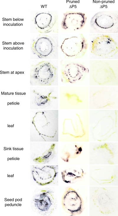
FIG. 5.
Immunoblots of various tissue sections from pruned or nonpruned S. sarrachoides plants infected previously with WT or the ΔP5 mutant at 9 weeks p.i. Areas positive for virus are indicated by purplish-blue staining. Virus levels and distribution were similar for pruned and nonpruned (not shown) plants infected with WT and QSS virus (pruned WT samples are shown). Virus levels and distribution for the SYG mutant (not shown) were similar to those of images shown for ΔP5. Similar to the images of healthy tissue shown in Fig. 3, no labeling was observed in healthy control tissue used in this experiment (not shown).

