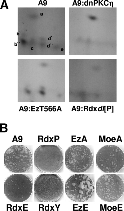
FIG. 7.
Impacts of ERM proteins on late stages of MVM infection. (A) A9 cells or derivatives thereof expressing dominant-negative PKCηT512A (A9:dnPKCη), dominant-negative Ez (A9:EzT566A), or dominant-negative Rdx (A9:Rdxdl[P]) were infected with MVM (30 PFU/cell) and 32P labeled with orthophosphate at 24 h p.i. Newly synthesized capsids were isolated by immunoprecipitation with αB7/αVP2 to enrich for DNA-containing virions and analyzed for the tryptic phosphopeptide pattern of VP2. (B) Impacts of ERM proteins on the capacity of MVM to form lysis plaques in parental A9 cells (A9) or derivatives expressing variant ERM proteins: Rdxdl[P] (RdxP), RdxT564E (RdxE), RdxY146F (RdxY), EzT566A (EzA), EzT566E (EzE), MoeT547A (MoeA), and MoeT547E (MoeE). No lysis plaques were detected in the presence of dominant-negative Rdxdl[P], while RdxY147F reduced plaque numbers 50-fold; in the remaining cell lines, plaque numbers were comparable to those of parental A9 cells.

