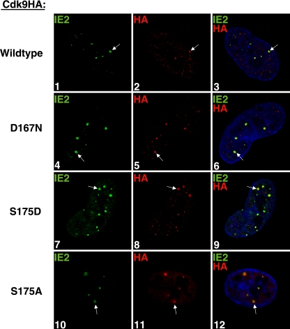
FIG. 5.
Neither cdk9 kinase activity nor binding to Brd4 is required for cdk9 localization at the viral transcriptosomes. G0-synchronized cells were released into G1, infected at an MOI of 5 with wt-cdk9HA HCMV (panels 1 to 3) or D167N-cdk9HA HCMV (panels 4 to 7), and seeded onto glass coverslips. The S175A-cdk9HA-expressing cells were induced with Dox as described in Materials and Methods. S175D- and S175A-cdk9HA cells were infected with HCMV Towne (MOI of 5) and seeded onto glass coverslips (panels 7 to 9 and 10 to 12, respectively). At 8 h p.i., cells were washed with PBS, fixed with 2% FA, permeabilized, and costained with antibodies specific for IE2 and HA (to detect exogenous cdk9HA). Fluorescein isothiocyanate- and tetramethylrhodamine isothiocyanate-conjugated isotype-specific secondary antibodies were used. Nuclei were stained with Hoechst dye. The white arrows point to examples of colocalization. All of the images represent confocal optical 0.2-μm sections at a magnification of ×1,000 under conditions of oil immersion.

