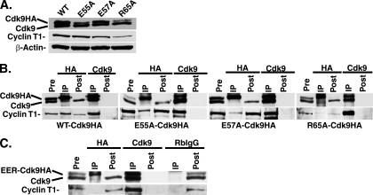
FIG. 6.
Amino acid changes in the N terminus of cdk9HA result in differential cyclin T1 binding. (A) The cell lines constitutively expressing wt-, E55A-, E57A-, and R65A-cdk9HA were lysed in 1× NETN buffer, prepared using 8% SDS-polyacrylamide gel electrophoresis (SDS-PAGE), and checked by Western blot analysis for the presence of cdk9 (endogenous and exogenous) and cyclin T1 protein. β-actin served as a loading control. (B) The cell lines constitutively expressing wt-, E55A-, E57A-, and R65A-cdk9HA were lysed in 1× NETN buffer and subjected to IP with antibodies for HA or cdk9. The IP, pre-IP, and post-IP lysates were prepared using 8% SDS-PAGE and checked by Western blot analysis for cdk9 and cyclin T1 protein. The E55A-cdk9HA and E57A-cdk9HA IP experiments were run on the same gel. The pre- and post-IP lanes represent 20% cell equivalents of the IP lanes. (C) The cell line expressing E55A/E57A/R65A-cdk9HA (EER-cdk9HA) was induced with Dox as described in Materials and Methods, lysed in 1× NETN buffer, and subjected to IP with antibodies for HA, cdk9, and an RbIgG control. The immunocomplexes (IP), pre-IP, and post-IP lysates were prepared using 8% SDS-PAGE and checked by Western blot analysis for the presence of cdk9 (endogenous and exogenous) and cyclin T1 protein.

