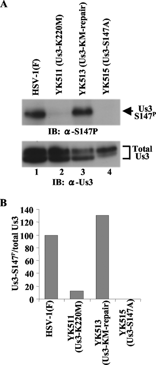
FIG. 3.
(A) Immunoblots of electrophoretically separated lysates from Vero cells infected with wild-type HSV-1(F) (lane 1), YK511 (Us3-K220M) (lane 2), YK513 (Us3-KM-repair) (lane 3), or YK515 (Us3-S147A) (lane 4). Infected Vero cells were harvested at 18 h postinfection and analyzed by immunoblotting with anti-Us3-S147P antibody (upper panel) or anti-Us3 antibody (lower panel). (B) Amount of Us3-S147P protein detected with anti-Us3-S147P antibody (A, upper panel) relative to the amount of Us3 protein detected with anti-Us3 antibody (A, lower panel) in HSV-1(F)-infected cells. The data were normalized to the value for HSV-1(F)-infected cells in panel A, lane 1. α, anti; Total Us3, Us3 protein detected by anti-Us3 polyclonal antibody.

