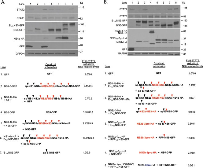
FIG. 3.
Expression of a precursor form of DENV NS5 cleaved by the DENV protease results in reduced STAT2 levels. (A) 293T cells were transfected with the indicated constructs. Twenty-four hours posttransfection, cells were sorted for GFP-positive cells by FACS, subsequently lysed, and examined via Western blotting using GFP-, HA-, STAT1-, STAT2-, and GAPDH-specific antibodies. Schematics of the transfected constructs are shown at the bottom. ORFs that contain an “sp” at the N terminus have a signal peptide which directs the entire polyprotein to the surface of the ER for translation. Black arrows and red arrows indicate cleavage sites for cellular and the DEN2 viral proteases, respectively. The DEN2 active protease is highlighted in red. Densitometry analysis of the levels of STAT2 and NS5 are included on the far right, and levels are calculated relative to the levels in lane 1, with a value of 1 in the case of NS5 levels being indicative of no detection (background levels). (B) Same as above (A). Mutation of the DENV protease recognition site at the N terminus of NS5 is indicated by the blue dashes. The DENV protease labeled in blue indicates a serine-to-alanine mutation within the catalytic site of the DENV protease.

