FIG. 1.
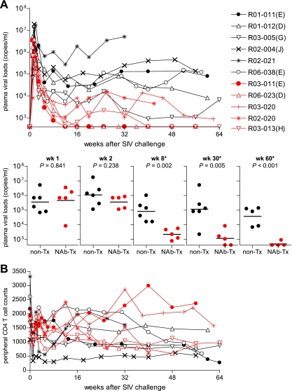
Follow up of NAb-immunized macaques. (A) Plasma viral loads (SIV gag RNA copies/ml plasma) in six unimmunized macaques (black lines) and five NAb-immunized animals (red lines) after SIVmac239 challenge. The plasma viral loads were measured as described previously (29). The lower limit of detection was approximately 4 × 10² copies/ml. The MHC-I haplotypes are shown in parentheses following the macaque numbers as follows: E, haplotype 90-010-Ie; D, 90-010-Id; G, 90-030-Ig; J, 90-088-Ij; and H, 90-030-Ih. Below are comparisons of plasma viral loads in unimmunized (non-Tx) and NAb-immunized (NAb-Tx) macaques at weeks (wk) 1, 2, 8, 30, and 60. The bars indicate the geometric mean of each group. The comparisons at weeks 8, 30, and 60 (indicated by asterisks) showed significant differences between two groups (P = 0.841 at week 1, P = 0.238 at week 2, P = 0.002 at week 8, P = 0.005 around week 30, and P < 0.001 around week 60 by t test). (B) Peripheral CD4+ T-cell counts (per μl) in unimmunized controls (black lines) and NAb-immunized macaques (red lines) after SIVmac239 challenge. The ratios of the counts around week 60 to those at week 0 in NAb-immunized macaques were significantly higher than in unimmunized controls (P = 0.028 by t test).

