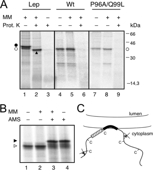
FIG. 2.
Full-length protein expression in vitro. (A) Proteinase K (Prot. K) treatment of microsomes carrying in vitro-translated WT Lep (lanes 1 to 3), the full-length PNRSV MP WT sequence (lanes 4 to 6), and mutant P96A/Q99L (lanes 7 to 9). Nonglycosylated and glycosylated molecules are indicated by the white dots and a black dot, respectively. An arrowhead indicates protease-protected fragments. MM, microsomal membranes. (B) The AMS derivatization of PNRSV MP. After translation, proteins were incubated with 20 mM AMS (+) or mock treated with dimethyl sulfoxide (−) in the presence (+) or absence (−) of microsomal membranes. (C) Model for the association of PNRSV MP with membranes; the engineered glycosylation sites and natural cysteines are cytoplasmically exposed.

