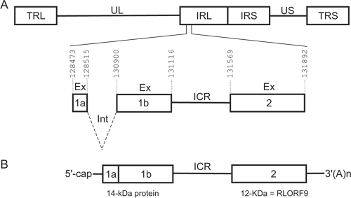
FIG. 1.
Genomic structure of MDV and positions of ORFs on the bicistronic transcript from the rightward transcriptional unit within the BamHI-H region. (A) Schematic representation of MDV genomic structure, consisting of unique long (UL) and unique short (US) regions, each bounded by a set of inverted repeats (TRL, IRL, IRS, and TRS). Intron (Int) and exon (Ex) sequences are shown, as well as the ICR between exon 1b and exon 2. (B) Schematic representation of the bicistronic transcript that we and others (21) cloned as cDNA. All genomic coordinates are according to the MDV-1 Md5 strain (GenBank accession number AF243438).

