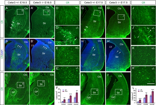
FIG. 3.
Accumulation of CR+ cells at the corticostriatal boundary and in the striatum of Celsr3 mutants. (A to E and A′ to E′) CR staining of the E18.5 subpallium. Large numbers of CR+ cells accumulate at the corticostriatal boundary (arrows in panels B and B′) at both rostral (in panels A and A′; counterstained with DAPI in panels B and B′), and caudal (C and C′) levels in Celsr3 mutants. Note the dilation of the lateral ventricle and the absence of axonal fibers (arrows in panel A) in the mutants (compare panels A′ to C′ with panels A to C). (D and D′) The accumulated CR+ cells have the morphology characteristic of migratory immature interneurons with leading processes in diverse directions (highlighted in insets), including dorsomedial (arrows), dorsolateral (empty arrowhead), and ventrolateral (arrowhead). (E and E′) Increased CR+ cells in the E18.5 Celsr3 mutant striatum. (F) Quantification of striatal CR+ cells demonstrates significant increases at the rostral, middle, and caudal levels. (G to K and G′ to K′) CR staining of the E17.5 subpallium. The accumulation at the E17.5 corticostriatal boundary is also obvious (compare panels J′ and D′). (L) The striatal CR+ cells are also increased in the E17.5 mutants throughout rostral, middle, and caudal levels. The data are in means ± standard deviations. A two-tailed Student's t test was used for statistics (n = 5): **, P < 0.01; ***, P < 0.001. Ctx, cortex; GP, globus pallidus; LV, lateral ventricle; Str, striatum. Bar, 500 μm (A to C, A′ to C′, G to I, G′ to I′) and 50 μm (D, D′, E, E′, J, J′, K, and K′).

