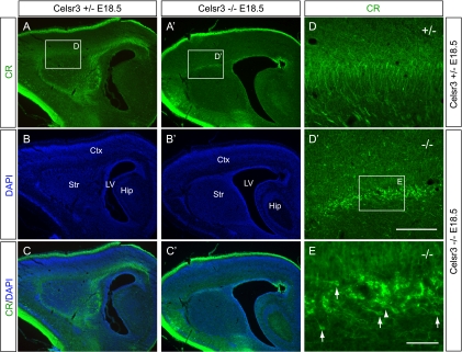
FIG. 4.
Sagittal view of the accumulated CR+ cells in the corticostriatal boundary. (A to E and A′ to D′) CR staining of the sagittal brain sections. The accumulated CR+ cells are migrating in multiple directions with leading process oriented rostrally (arrows in panel E) or caudally (arrowhead in E). Ctx, cortex; Hip, hippocampus; LV, lateral ventricle; Str, striatum. Bar in panel D′, 800 μm for panels A to C and A′ to C′ and 200 μm for panels D and D′; bar in panel E, 50 μm.

