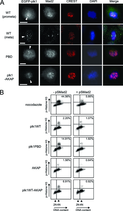
FIG. 6.
Spindle checkpoint activation in Plk1-AKAP-expressing cells. (A) Mad2 remains associated with kinetochores in Plk1-AKAP expressing cells, U2OS cells were transfected with the indicated constructs as in Fig. 4, and released at 20 h after thymidine block. Cells were fixed and stained with anti-Mad2 antibody, CREST, and DAPI. Images were collected and processed using OpenLab (Improvision) or a Deltavision microscope system (Applied Precision). Plk1 at mitotic centrosomes is indicated by arrowheads. Merged images show Mad2 in green, CREST staining in red, and DAPI in blue. All scale bars represent 5 μm. (B) U2OS cells were transfected with the indicated EGFP-tagged Plk1 plasmids with or without an shRNA-expressing vector targeting Mad2 (pS-Mad2). At 20 h after release from a thymidine block, cells were harvested and fixed for FACS analysis. In the control experiments examining spindle checkpoint activation by nocodazole, cells were transfected with pS vector control or pS-Mad2, along with spectrin-GFP, and nocodazole was added for 20 h after release of the thymidine block (top panels). Phospho-histone H3 positivity and DNA content (PI) of GFP-positive cells are shown.

