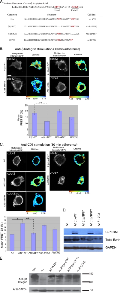
FIG. 4.
NPIY motif of β1 integrin is important for Cdc42 activation. The β1 integrin-deficient A1 cells and the various A1-derived lines that have been reconstituted with the WT and mutated β1 integrin constructs (sequences summarized in panel A, where …… depicts the deleted regions) were adhered on anti-β1 integrin (12G10) (B) or anti-CD3 (UCHT1) (C) antibody-coated coverslips for 30 min at 37°C. This time point was chosen on the basis of the maximum difference between anti-β1- and anti-CD3-induced Cdc42 activities observed from the data in Fig. 1D. Bar charts represent the cumulative FLIM/FRET data of three experiments (n = 7 cells) (*, P < 0.05; **, P < 0.005). Scale bar = 5 μm. (D) Western blot analysis of whole-cell lysates from A1 cells and the various β1 integrin-reconstituted A1-derived lines. Blots were probed with anti-C-PERM (ERM phosphorylated at the conserved threonine residue in the COOH terminus) rabbit IgG (Cell Signaling) or anti-total ezrin (an in-house MAb, 2H3) antibody as described before (53). (E) Western blot analysis of whole-cell lysates from A1 cells and the various β1 integrin-reconstituted A1-derived lines. Blots were probed with an anti-total β1 integrin antibody (MAb 8E3; a kind gift of Martin Humphries, University of Manchester). Results shown are representative of two independent experiments.

