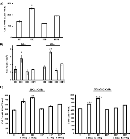
FIG. 1.
PRL attenuates EGF-induced cell proliferation of mammary epithelial cells. (A) HC11 cells (1.5 × 10³ cells) were plated in assay media (2% FBS, HI) in the absence or presence of PRL (1 μg/ml) o/n. Cells were then left untreated or treated with EGF (10 ng/ml) for 72 h. MTT assays were performed as described in Materials and Methods. Results are the means ± SEM for triplicates of three experiments (*, P = 0.043). (B) HC11 cells (2.5 × 10⁴ cells) were plated as described for panel A, and cells were then left untreated or treated with EGF for 24 h and 48 h. Cell numbers were determined following trypan blue exclusion assays. Results are the means ± SEM for triplicates of three experiments (*, P = 0.07; **, P = 0.049). (C) HC11 cells (1.5 × 10³ cells) (left panel) and NMuMG cells (3 × 10³ cells) (right panel) were plated as described for panel A, and cells were then left untreated or treated with either 10 ng/ml or 100 ng/ml EGF (E-10ng and E-100ng, respectively) for 72 h. MTT assays were performed, and results are the means ± SEM for triplicates of three experiments (*, P = 0.0189; **, P = 0.0165; ***, P = 0.002; ****, P = 0.0004). Abs, absorbance.

