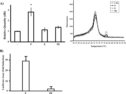
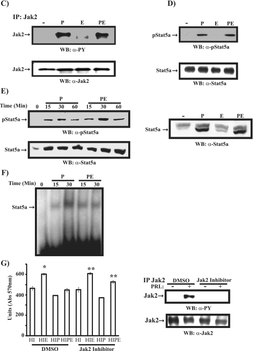
FIG. 3.
EGF regulation of PRL-induced Jak2/Stat5a pathway and β-casein gene expression. (A, left) Differentiated HC11 cells were left untreated or treated with PRL (P), EGF (E), or PRL/EGF (PE) for 16 h. qRT-PCR of the β-casein gene was performed, and results are the means from three experiments (*, P = 0.053). (Right) Dissociation curve analysis of the q-PCR. The four identical peaks confirm that the primers for the β-casein gene are specific, resulting in one amplified field. CTL, cytotoxic T lymphocytes. (B) Differentiated HC11-Lux cells were left untreated or treated with either PRL or PRL/EGF for 16 h. Results are the mean luciferase activity levels from quadruplicates of two experiments. (C) Serum-starved differentiated HC11 cells were untreated or treated with PRL, EGF, or PRL/EGF for 15 min. Cell lysates were immunoprecipitated (IP) with a polyclonal antibody to Jak2, immunoblotted with a monoclonal antibody to phosphotyrosine (upper panel), and reprobed with a polyclonal antibody to Jak2. (D) Cell lysates described for panel C were immunoblotted with a monoclonal antibody to phospho-Stat5a (upper panel) and reblotted with a monoclonal antibody to Stat5a (lower panel). (E, left) HC11 nuclear extracts were immunoblotted with a monoclonal antibody to phospho-Stat5a (upper panel) and Stat5a (lower panel). (Right) Nuclear extracts of HC11 cells treated with PRL, EGF, or PRL/EGF for 30 min were immunoblotted with a monoclonal antibody to Stat5a. (F) EMSA was performed using the Stat5a binding site of the β-casein gene promoter with nuclear extracts prepared as in panel E. (G, left) HC11 cells were treated with either DMSO or Jak2 inhibitor (1 μM) and grown o/n in 2% serum containing HI or HIP. Cells were then left untreated or treated with EGF for 48 h. An MTT assay was performed, and results are the means ± SEM for triplicates of three experiments (*, P = 0.017; **, P = 0.023). (Right) HC11 cells were pretreated with either DMSO or Jak2 inhibitor (25 μM) for 2 h. Cells were then stimulated with PRL for 15 min. Cell lysates were immunoprecipitated with a polyclonal antibody to Jak2 and immunoblotted with a monoclonal antibody to phosphotyrosine (PY) (upper panel) and reprobed with a polyclonal antibody to Jak2 (lower panel). Abs, absorbance; WB, Western blot; α-, anti-.


