FIG. 5.
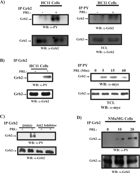
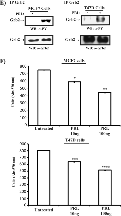
PRL induces tyrosine phosphorylation of Grb2 in mammary cells. (A, left) Serum-starved HC11 cells were untreated or treated with PRL for 15 min. Lysates were immunoprecipitated (IP) using a polyclonal antibody to Grb2 and immunoblotted with a monoclonal antibody to phosphotyrosine (upper panel) and a polyclonal antibody to Grb2 (lower panel). (Right) Lysates of HC11 cells prepared as described above were immunoprecipitated using a polyclonal antibody to phosphotyrosine and immunoblotted with a polyclonal antibody to Grb2 (upper panel). Cell lysates were immunoblotted with a polyclonal antibody to Grb2 (lower panel). (B, left) Serum-starved differentiated HC11 cells were pretreated with sodium vanadate (50 μg/ml) for 30 min and then stimulated with PRL for 15 min. Lysates were immunoprecipitated with a polyclonal antibody to Grb2, immunoblotted with a monoclonal antibody to phosphotyrosine (upper panel), and reblotted with a polyclonal antibody to Grb2 (lower panel). (Right) Differentiated HC11 cells were transfected with an expression vector encoding myc-Grb2. Serum-starved cells were pretreated with sodium vanadate (50 μg/ml) for 30 min, stimulated with PRL as indicated, and immunoprecipitated using a polyclonal antibody to phosphotyrosine, followed by immunoblotting with a monoclonal antibody to the myc tag (upper panel). Lysates from the same transfection were immunoblotted with a monoclonal antibody to the myc tag (lower panel). (C) Serum-starved differentiated HC11 cells were pretreated with DMSO or Jak2 inhibitor (25 μM) for 90 min. Cells were then left unstimulated or were stimulated with PRL for 15 min. Lysates were immunoprecipitated and immunoblotted as in panel A (left). (D) Serum-starved NMuMG cells were left untreated or were treated with PRL as indicated. Numbers indicate time (in minutes). Lysates were immunoprecipitated using a polyclonal antibody to Grb2 and immunoblotted with a monoclonal antibody to phosphotyrosine (upper panel) and a polyclonal antibody to Grb2 (lower panel). (E) Serum-starved MCF-7 (left panel) and T47D (right panel) cells were pretreated with sodium vanadate (50 μg/ml) for 30 min and then stimulated or not with human PRL (100 ng/ml). Cell lysates were immunoprecipitated with a polyclonal antibody to Grb2, immunoblotted with a monoclonal antibody to phosphotyrosine (upper panel), and reblotted with a polyclonal antibody to Grb2 (lower panel). (F) MCF-7 (upper panel) and T47D (lower panel) cells were plated o/n in 2% serum and left untreated or treated with human PRL for 72 h. An MTT assay was performed, and the results are the means ± SEM for triplicates of three experiments. Upper panel, *, P = 0.0216; **, P = 0.0075. Lower panel, *, P = 0.0153; **, P = 0.0024. Abs, absorbance; WB, Western blot; α-, anti-; PY, phosphotyrosine; TCL, total cell lysates.


