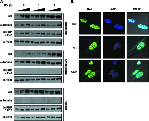
FIG. 5.
(A) IMR-90 cells were treated with NO for the times shown, whereupon whole-cell, cytoplasmic, and nuclear lysates (2.5, 5.0, and 7.5 h) were prepared and the levels of HuR, the cytoplasmic marker α-tubulin, the nuclear marker hnRNP C1/C2, and the loading control β-actin were determined. (B) Immunofluorescence analysis of HuR. HuR signals (green) in either untreated (Unt.) or NO-treated (0.5 mM for 4 h) IMR-90 cells were studied. Green, HuR fluorescence; blue, DAPI staining to visualize nuclei; merge, overlap of the two signals. Treatment of IMR-90 cells with CGP74514A (2 μM for 2 h), which induces HuR translocation to the cytoplasm, was included as a positive control.

